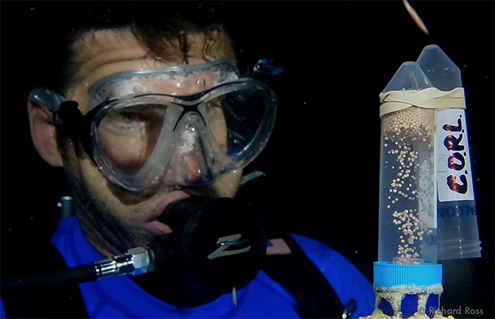
TOMLOV DM602 is the Perfect All-Around Microscope for Reefkeeping
From ReefBuilders
Microscopes are cool and have lots of uses for reefkeeping: looking at the critters in the sand, looking at the details of a coral’s surface, trying to ID pests, and more. But picking a microscope can be daunting. There are a million microscopes available that cost anywhere from about 70 dollars and into the thousands of dollars. So, what do you pick? A compound microscope? A dissecting microscope? Stereo or monocular? Built-in camera? Digital screen? It all depends on what you are going to use the microscope for.
But, what if you want to do everything from look at Acro Eating Flatworms and their eggs, examine gill and fin clips or skin scrapes, identify strains of dinoflagellates, see what is living in your sand, or care for baby corals? Well, I just got a new microscope that changed my world, and it may change yours as well for viewing, photographing, filming, and working on reef tank life – at $260 (USD), it won’t break the bank.
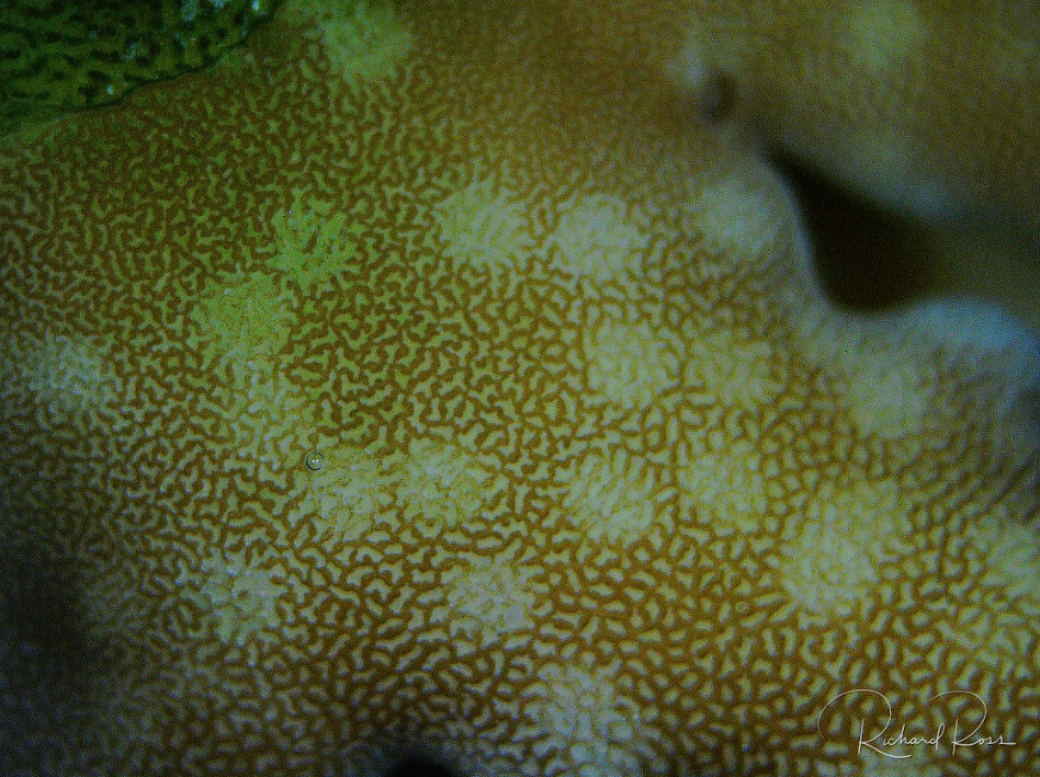 AEFW bites on an Acropora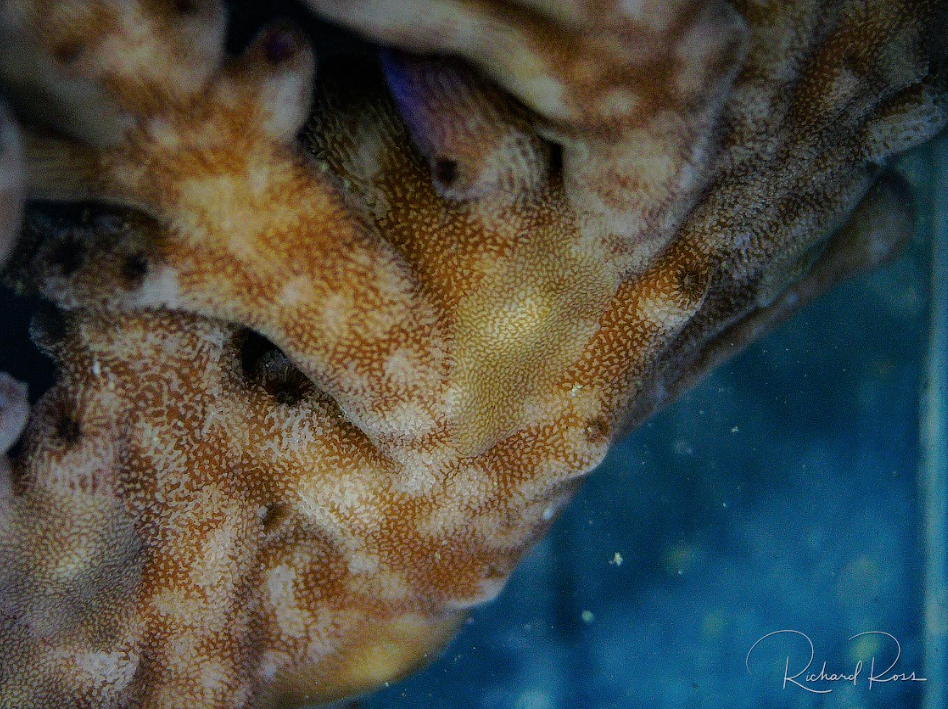
The issues
For the Acropora spawning work I do, I use a microscope constantly. For the first few days after the spawn and the fertilization of embryos, folks spawning corals use microscopes to see if fertilization has occurred and to track how well the embryonic corals are developing. After the embryos become larvae and then settle onto a substrate, the real work with the microscope begins. Basically, everything wants to kill baby corals. Algae and coralline algae can smother baby corals in a matter of days, while evil stuff like hydroids can pop up and sting the babies into sadness overnight. Not only do you need to be able to visualize what is happening in the vicinity of a single coral polyp less than a millimeter in diameter, but you also need to be able to use tiny tools to scrape, pull, and pluck baby coral killers while keeping the coral alive.
Dissecting scopes are used to visualize the surface features of an object, while compound microscopes are used to look at objects on a cellular level. For most live coral work, we are interested in the surface features, and we don’t want to destroy what we are looking at by preparing it to go on a microscope slide with a cover slip as we are essentially using a dissecting scope as a macro lens. I have been using a dissecting scope for this work for years, but the staging areas are small and cramped because they are made for visualization, not scraping algae with dental tools.
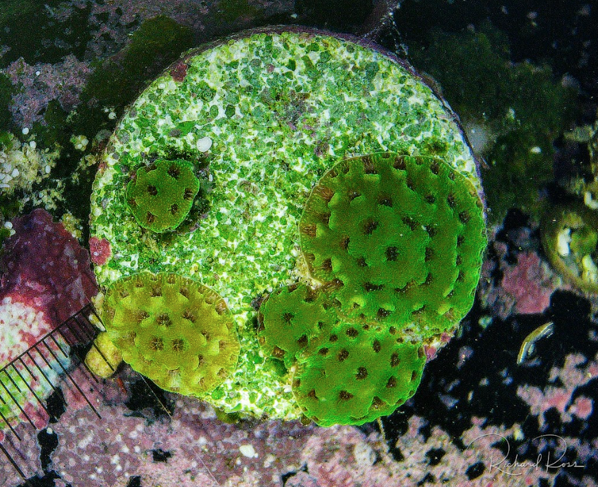 Four month old Acropora millepora.
Part of the problem with the small staging area of a traditional microscope is that living coral can’t be put on a microscope slide and be expected to stay alive. The living coral needs to stay submerged in saltwater while we examine its skin, structure, and whatever is living on the coral. Aquarium people are clever folks and find or build containers that can ‘make it work’, but even then, the size of the coral that can be examined is extremely limited, as well as the angles the microscope can be used to visualize the animal.
Staring into the eyepieces of a microscope while working for hours can get frustrating fast. Even more annoying, taking pictures on these scopes is often harder than we want it to be with many budget digital microscopes having very low-quality imaging output. I have taken to using a scope mount for my iPhone over a microscope-based photography solution that produces great results but is a bit cumbersome to set up and use.
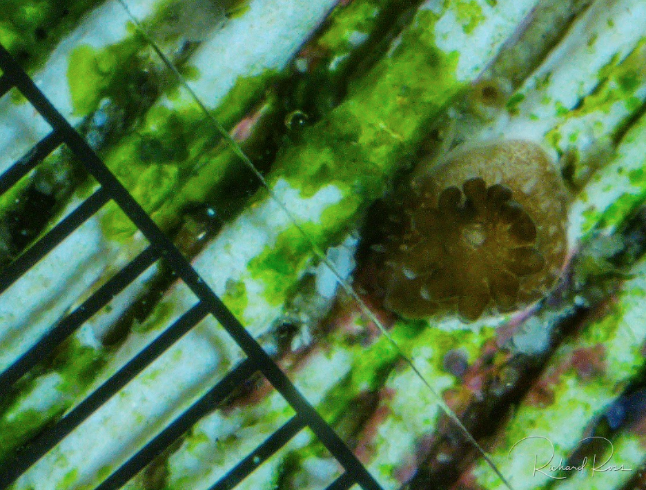 Newly settled P. damicornis.
I bought an affordable dissecting scope for last year’s spawn and still ended up chopping it up with a Dremel tool so the staging area was big enough for me to do the work I needed to do, and even then, it was still cramped. I also had to buy a set of microscope lights to light up the baby coral from different angles, because the built-in top and bottom lights don’t effectively light up larger items under the scope. The scope, lights, and iPhone mount together cost me about 200 dollars and it was still sub-optimal. There has got to be a better way!
The Better Way
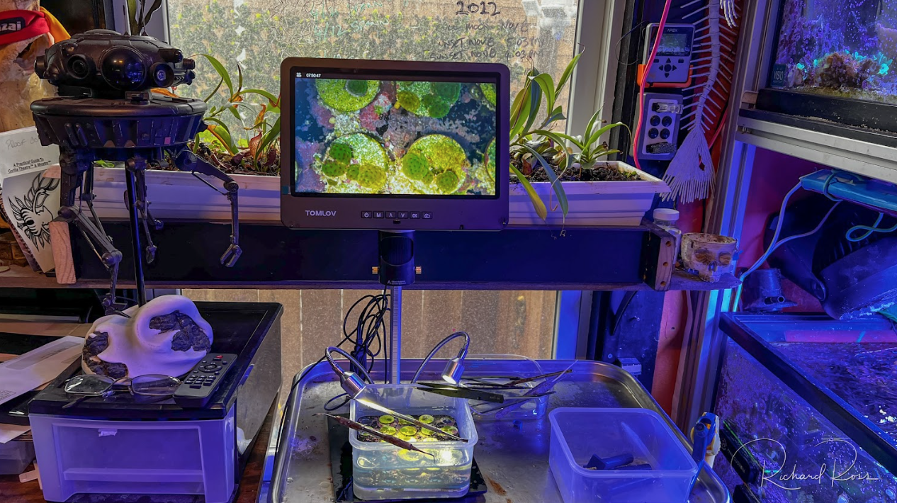
The TOMLOV DM602 HDMI Digital soldering microscope is used primarily for soldering electronics and by coin collectors to inspect coins. Instead of eyepieces, this baby has a 10.1-inch digital screen that has a sharp picture and is positionally adjustable, making long microscope work sessions so much easier on the neck and back. It also has built-in dimmable lights on flexible stalks to make side lighting easier and more effective. Even better, the staging area has no built-in under-lighting or slide clips so there is so much more room to work (included in the box is a removable stage with built-in under-lighting for slide work). TOMLOV even makes an arm bracket that mounts to a desk, eliminating the stage altogether so there is nothing at all under the scope to interfere with the workspace. All in all, it’s just perfect for the work I am doing with baby coral.
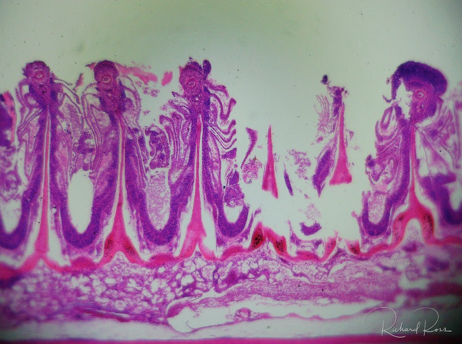 A prepared fish gill clip.
The scope comes with a three-lens kit that is easy and fast to swap out, so this scope is good for anything you might want to look at from your reef tank. If you want to look at gill clippings, try to ID dinos, look at tiny critters in the sand or water, or visualize coral polyps, you are all set. Video and photo quality is amazing for the price of ($)269 USD, so for most of us, this scope is an all-around win.
The TOMLOV DM602 HDMI Digital Microscope currently sells on Amazon for ($)269. There are many other makes and models of soldering microscopes that have a digital screen and look like they should work well, so if you find one that takes better pics or video, please let me know. I also imagine that it is only a matter of time before these kinds of units become even better and even less expensive.
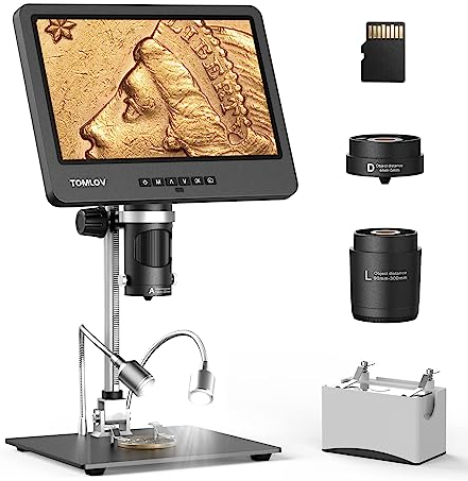
The TOMLOV DM602 with its 10.1-inch screen is a great all around magnification solution for the reef aquarist, allowing us to examine living coral as well as prepared samples on microscope slides for disease and pest identification at a cellular level. The video quality is excellent, and the photo quality is very good. For $269 USD, it is the perfect all-around microscope solution for the reef hobbyist.
About the author
Rich Ross is the recipient of the MASNA Aquarist of the Year award, an independent coral spawning and cephalopod researcher, and co-host of the Reef Beef Podcast. See and hear more from him when he appeared as a guest on the Reef Therapy Podcast.

Richard is known for his “Skeptical Reefkeeping” article series, his one-of-a-kind presentations, his coral spawning and cephalopod research, the Reef Beef Podcast, and for managing the 212,000 gallon reef tank in the Steinhart Aquarium at the California Academy of Sciences. He has kept saltwater animals and tanks for over 40 years, and has won multiple awards including the 2014 MASNA Aquarist of the Year. His work has been covered by Scientific American, National Geographic, Animal Planet, Penn’s Sunday School, NPR’s Science Friday, Discovery News, Adam Savage, Fox News, and more.